BIU scientists Pioneer Use of Worms Rather than Rats to Model Human Muscle Diseases
Instead of testing rodents that display species-related differences and present ethical and budgetary concerns, Bar-Ilan University researchers develop innovative new method

Conducting health research on animal models is vital for promoting biological and medical studies and allow analysis of molecular and pathophysiological pathways of diseases and pathological conditions. Such models make possible the repurposing of approved drugs for conditions for which they were not originally intended and the development of novel therapeutics.
Rodents are the most common models of human disease, but studies using these models are hampered by species-related differences and ethical and budgetary concerns – so alternative models, tightly related to human physiology and disease pathologies are needed.
Now, researchers from BIU have developed a novel platform to model human muscle diseases in the Caenorhabditis elegans (C. elegans) worm. The species is a small nematode about one millimeter long used as a model non-human organism to help scientists understand biological processes. The innovation promotes the study of diseases in a versatile, scalable way, opening the door to more personalized approaches to disease modeling.
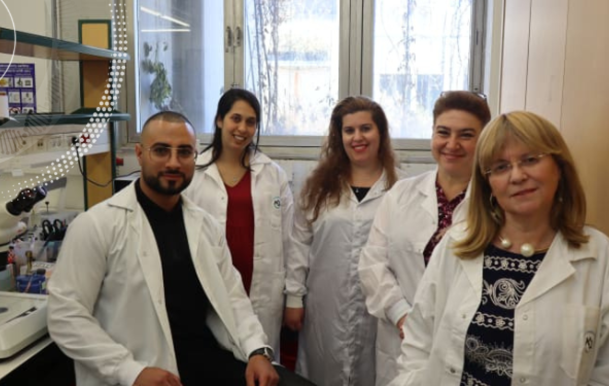
The research team was led by Prof. Chaya Brodie, from BIU’s Goodman Faculty of Life Sciences and Institute of Nanotechnology and Advanced Materials (BINA), and Prof. Sivan Korenblit, from the Goodman Faculty, in collaboration with Dr. Amir Dori, a muscular dystrophy specialist from Sheba Medical Center at Tel Hashomer. They have just published their study in the journal Disease Models and Mechanisms under the title “Cross-species modeling of muscular dystrophy in Caenorhabditis elegans using patient-derived extracellular vesicles
They harvested extracellular vesicles (cell-derived vesicles surrounded by membranes that carry bioactive molecules and deliver them to cells) from patients with Duchenne muscular dystrophy and transferred them to C. elegans worms. The results were remarkable, with the worms developing muscle atrophy very similar to human symptoms.
This achievement was made possible by a novel approach that utilizes extracellular vesicles derived from blood samples taken from patients. “Unlike traditional methods that rely on genetic modifications in model organisms, this technique relies on elements secreted into the blood,” explained doctoral student Rewayd Shalash, who carried out the research with doctoral student Coral Cohen and Dr. Mor Levi-Ferber.
Originally published on The Jerusalem Post.